 |
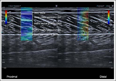
Figure 2.
Representative shear-wave elastography (SWE) image of the medial gastrocnemius showing five regions of interest (ROIs) defined from superficial to deep: deep fascia (DF), superficial portion of the medial gastrocnemius (UMG), middle portion (MMG), deep portion (LMG), and deep intermuscular fascia (DIF). The color map indicates shear modulus, with warmer colors representing higher values (greater stiffness).