Research article - (2025)24, 578 - 588
DOI:
https://doi.org/10.52082/jssm.2025.578
Enhancing Jump Performance Through Blood Flow Restriction Squat Exercise: A Muscle Synergy Analysis Using Wavelet Packet Transformation
Chenxi Hu1,2,, Ning Du3,1,, Yanfeng Li2, Olivier Girard4, Tao Mei1,5,
1China Institute of Sport and Health Science, Beijing Sport University, Beijing, China
2Institute of Artificial Intelligence in Sports, Capital University of Physical Education and Sports, Beijing, China
3Department of Orthopaedics, Fourth Medical Centre of Chinese PLA General Hospital, Beijing, China
4School of Human Sciences (Exercise and Sport Science), The University of Western Australia, Crawley, Western Australia
5Deqing Alpha Innovation Institute, University of Science and Technology of China, Hangzhou, Zhejiang, China
† The authors have the same contribution and thus share the role of the first author

Tao Mei
✉ China Institute of Sport and Health Science, Beijing Sport University, Beijing, 100084, China
Email: meitao@bsu.edu.cn
Received: 04-05-2025 -- Accepted: 29-06-2025
Published (online): 01-09-2025

ABSTRACT

To explore neuromuscular control during blood flow restriction (BFR) squat exercise using wavelet packet transform (WPT) combined with non-negative matrix factorization (NMF). Fifteen resistance-trained males completed four sets of squats at 40% arterial occlusion pressure. Countermovement jump (CMJ) height and reactive strength index modified (RSImod) alongside surface electromyographic activity from eight lower-limb muscles were assessed before after the exercise. CMJ height and RSImod significantly increased post-exercise (P < 0.001, Cohen's d = 0.45 and 0.34, respectively). Four muscle synergy modules were consistently identified, though primary muscle contributions shifted across movement phases. The tibialis anterior (TA) was the primary contributor in Synergy1, while the gastrocnemius lateralis (GL) dominated Synergy 2, accompanied by a significant increase in gluteus maximus (GM) weight (P = 0.032). In Synergy 3, the rectus femoris (RF), vastus lateralis (VL), and vastus medialis (VM) were predominant, with significant changes in GM and VM muscle weights (P = 0.013, 0.039). Synergy 4 was characterized by contributions from the semitendinosus (ST), biceps femoris (BF), and GM, with a significant increase in VL muscle weight (P = 0.024). WPT-NMF analysis revealed distinct time-frequency synergy modules in CMJ movements before and after BFR squat exercise. Significant changes in activation weights were observed within the 0-250 Hz range (P < 0.05). BFR squat exercise acutely enhances countermovement jump performance by refining muscle synergy and neuromuscular activation patterns, providing novel insights into neuromuscular control strategies.

Key words: Blood flow restriction training, countermovement jump, muscle synergy, wavelet packet transform, non-negative matrix factorization

Key Points
  • Wavelet packet transform combined with non-negative matrix factorization effectively captures time-frequency muscle synergy patterns, offering new insights into neuromuscular control during countermovement jump post-BFR exercise.
  • Acute BFR exercise enhances countermovement jump performance by optimizing muscle activation patterns, without altering the number of synergy modules.
  • Frequency-specific shifts in muscle activation suggest that the central nervous system modulates synergy through adaptive, frequency-dependent control strategies to optimize explosive strength.
  • BFR exercise serves as an effective strategy to maximize lower-limb power, with direct applications in sports requiring explosive movements.
INTRODUCTION

Blood flow restriction (BFR) involves applying a cuff or tourniquet to the proximal portion of a limb to limit venous return, while partially restricting arterial inflow. This technique typically involves moderate external limb compression (40-60% of arterial occlusion pressure) during low-load exercises such as squats, which are commonly used in BFR training protocols (Lorenz et al., 2021). By reducing mechanical load while increasing physiological stress, BFR training offers an effective low-load alternative to traditional heavy resistance exercise, with strength gains comparable to heavier loads (Wortman et al., 2021).

The countermovement jump (CMJ) is a widely used test of vertical jump performance, reflecting lower limb explosive power.(Ramirez-Campillo et al., 2022). BFR resistance exercise can potentiate CMJ performance, including vertical jump height and rate of force development (RFD). For instance, half-squat exercises performed at 50-70% arterial occlusion pressures led to a 7% improvement in CMJ height in female soccer players (Sun and Yang, 2023). Similarly, BFR added to leg half-squats improved CMJ height under varying external loads in volleyball players (Wang et al., 2022). Squats are widely used in BFR protocols due to their ability to activate multiple lower-limb muscle groups, making them particularly effective for strength and power development (Cornejo-Daza et al., 2025; Jagim et al., 2024; Wang et al., 2022). Given the biomechanical similarity between squats and CMJ, examining the impact of BFR squat training on CMJ performance can provide valuable insights into underlying neuromuscular adjustments. Since CMJ engages multiple joints and muscles, investigating muscle synergies offers a novel lens on how BFR enhances jump performance.

Muscle synergy analysis is a method for studying neuromuscular control by revealing how the central nervous system (CNS) coordinates multiple muscles through spatiotemporal activation patterns (Beltrame et al., 2023). This approach has demonstrated cross-task stability and broad applicability, enabling detailed examination of weight distribution, temporal features, and control pattern variations in motor coordination (Bao Nguyen et al., 2017; Goudriaan et al., 2022). Muscle synergy analysis is increasingly applied in sports skill assessment, injury mechanism analysis, and rehabilitation monitoring, reflecting a shift from single-muscle analysis toward a more integrated, system-level understanding of neuromuscular control (Turpin et al., 2021).

Non-negative matrix factorization (NMF) is widely used in synergy analysis to decompose multi-dimensional surface electromyography (sEMG) signals into distinct synergy patterns. By preserving non-negativity in both the decomposed matrices and basis vectors, NMF aligns with the inherent characteristics of sEMG signals and improves interpretability (Xie et al., 2022). However, NMF has limitations in capturing time-varying characteristics, as it decomposes signals into fixed spatial and temporal components. This poses challenges in dynamic sEMG analysis, where different frequency bands encode complex physiological information related to motor control (Vigotsky et al., 2017). Consequently, NMF is less effective in contexts requiring detailed time-frequency analysis to fully understand complex neuromuscular processes.

To address the limitations of traditional NMF, wavelet packet transform-based non-negative matrix factorization (WPT-NMF) has been introduced to capture both time- and frequency-dependent characteristics of EMG signals during dynamic movements (Xie et al., 2022). The CMJ, which involves rapid muscle activation and motor unit recruitment, contains high-frequency sEMG components indicative of complex motor unit recruitment information (Enoka, 2008). Therefore, time-frequency-based muscle synergy analysis using WPT-NMF is particularly well-suited for studying dynamic tasks like CMJ. Thus, WPT-NMF enables detailed synergy analysis in explosive tasks like CMJ. A previous study by our group applied WPT-NMF to examine muscle synergies during BFR squat exercise under varying external compression parameters, and found that pressure differences selectively influenced the number of synergies within specific frequency bands (Chang and Hu, 2025). Building upon this framework, the present study shifts focus to pre–post intervention changes during CMJ, aiming to investigate acute neuromuscular adaptations induced by BFR exercise.

While BFR exercise is widely recognized for enhancing athletic performance, its impact on muscle synergy during CMJ execution remain underexplored. To address this gap, we collected EMG signals during CMJ tasks performed before and after an acute BFR squat session and analyzed time-frequency muscle synergies using the WPT-NMF method. We hypothesized that acute BFR squat exercise would improve CMJ performance and alter time-frequency muscle synergy patterns. Specifically, we expected trained males to exhibit adjustments in muscle synergies across frequency bands during CMJ execution post-intervention, reflecting neuromuscular adjustments associated with BFR-induced performance improvements.

METHODS
Participants

Fifteen healthy male participants (age: 23.4 ± 2.2 years; height: 178.5 ± 5.4 cm; body weight: 76.7 ± 7.5kg; 1RM: 139.4 ± 21.8kg) were recruited. Participants met the criteria for the Trained/Developmental level, as defined by McKay et al. (2022), based on their resistance training history and movement proficiency. Inclusion criteria were: (1) at least three years of resistance training experience, (2) ability to perform a 1-repetition maximum (1RM) squat exceeding 1.5 times their body weight, and (3) proficiency in both CMJ and squat techniques. Exclusion criteria included: (1) current or recent musculoskeletal or joint injuries involving either the upper or lower limbs (within the past six months), (2) diagnosed cardiovascular conditions, and (3) any other medical conditions that could contraindicate participation in high-intensity lower-limb exercise. The study was approved by the Ethics Committee of Capital University of Physical Education and Sports (Approval No. 2025A074), in accordance with the Declaration of Helsinki. Because of the nonlinear nature of muscle synergy data, traditional power analysis using G*Power may not be appropriate(Guo et al., 2025). Prior studies using similar synergy analysis included 10 to 14 participants (Fan et al., 2024; Matsunaga et al., 2021). Based on the similarity in methodology and task features, the present study included 15 participants.

Experimental procedure

The study consisted of two phases: a preparation phase and an exercise intervention phase. During the preparation phase, participants' age, height, weight, 1RM, and thigh circumference were recorded to calculate individual training loads and occlusion pressures. In the intervention phase, participants first completed a 10-minute running warm-up, followed by the placement of sEMG electrodes and BFR cuffs. They then performed three CMJ trials following standardized testing protocols (Sun and Yang, 2023). The two highest jumps (based on jump height) were selected for further analysis, and the corresponding EMG signals were used for muscle synergy analysis. After CMJ testing, participants rested for one minute before performing four sets of occluded squats. During the BFR squat session, participants performed four sets of occluded squats, with 1-minute inter-set rest intervals, resulting in approximately 4 minutes of accumulated recovery. Post-exercise CMJ testing was conducted immediately following the final set, without additional rest. Throughout the experiment, CMJ performance and EMG signals were synchronously collected using a force platform (Kistler, USA) and EMG equipment (Delsys, USA). The full experimental procedure lasted approximately 60~75 minutes per participant, and all testing was performed between 2:00 p.m. and 5:00 p.m. to minimize circadian variation.

Exercise intervention

The exercise intervention consisted of resistance squats performed on a squat rack, following a standardized low-load exercise routine at 30% of each participant’s 1RM. The protocol included four sets: the first set consisted of 30 repetitions, followed by three sets of 15 repetitions, with 60 seconds of rest between sets (Patterson et al., 2019). Squat tempo was standardized to 2 seconds for the eccentric (downward) phase and 1 second for the concentric (upward) phase (Patterson et al., 2019).

Occlusion pressure was applied using a pneumatic cuff and occlusion pump (Harvard Apparatus, USA), starting 5 seconds before each set. The cuff was positioned just below the gluteal fold, perpendicular to the thigh (Patterson et al., 2019). After the pressure was applied, participants began the first squat set. The cuff was deflated during rest periods and re-inflated 5 seconds before the next set. This cycle was repeated for all four sets, with the cuff was removed after the final set.

Individual occlusion pressure was determined based on thigh circumference and the arterial occlusion pressure estimation method described elsewhere (Loenneke et al., 2015), with the target pressure set at 40% of each participant’s arterial occlusion pressure (Loenneke et al., 2015). An overview of the entire experimental procedure, including EMG application, testing, and BFR intervention, is illustrated in Figure 1.

Measurements

1RM Test

Participants first completed a 10-minute treadmill warm-up, followed by 5-10 repetitions at 40% to 60% of their estimated 1RM. After a 1-minute rest, they performed 3-5 repetitions at 60-80% of their estimated 1RM. For the formal testing phase, participants began with a load set at 90% of their estimated 1RM. If successful, the load was progressively increased (Comfort et al., 2018). Attempts were separated by 3~5 minutes, with participants deciding when to proceed based on their perceived recovery. If an attempt failed, the last successfully lifted load was recorded as the 1RM (Kraemer and Ratamess, 2004). All participants achieved their 1RM within five attempts. The test was supervised throughout by two trained researchers who provided verbal encouragement. The protocol was terminated if the attempt failed or if proper squat technique could not be maintained (Oláh et al., 2025).

Countermovement jumps

The CMJ test was conducted on a three-dimensional force platform with a sampling frequency of 1000 Hz. The testing procedure was as follows: (1) Participants stood on the force platform in a stationary position with hands placed on their hips, and (2) On the tester's command, they initiated a countermovement by flexing their knees, then jumped vertically as high as possible, followed by controlled landing. CMJ height and the reactive strength index modified (RSImod) were calculated from the vertical force trajectory recorded during each jump (Healy et al., 2018). The CMJ movement was divided into five distinct phases (Figure 2): unweighting, braking, propulsion, flight, and landing. Test-retest reliability was assessed using ICC(2,1), with values of 0.95 and 0.96 for CMJ height and 0.71 and 0.81 for RSImod before and after the intervention, respectively.

Lower limb sEMG testing

A 16-channel wireless sEMG system was used to monitor signals from eight lower-limb muscles: rectus femoris (RF), vastus lateralis (VL), vastus medialis (VM), semitendinosus (ST), biceps femoris (BF), gluteus maximus (GM), tibialis anterior (TA), and gastrocnemius lateralis (GL). The system sampled at 2000 Hz and was synchronized with the Kistler three-dimensional force platform.

Before each test, the sEMG acquisition system was calibrated and set to standby mode. Testing commenced after participants completed their warm-up, with sEMG signals recorded during the CMJ trials following the established protocol. The dominant frequency range of the sEMG signals was 0-250 Hz. The time-frequency analysis revealed higher amplitudes primarily within the 250Hz band (Figure 3). Therefore, the frequency range studied was 0-250 Hz, divided into four bands: 0-62.5 Hz, 62.5-125 Hz, 125-187.5 Hz, and 187.5-250 Hz.

Data analysess

EMG signal pre-processing

For the NMF-based muscle synergy analysis, the following steps were taken: 1) A second-order Butterworth notch filter was applied to remove 50 Hz powerline interference; 2) A second-order Butterworth band-pass filter (20-400 Hz) was applied to isolate the target frequency band signals; 3) The filtered signals were full-wave rectified; 4) The RMS envelope was extracted and normalized to the maximum value; 5) The envelope signals were time-normalized to 0-100%. For WPT-NMF muscle synergy analysis, steps 3, 4, and 5 from the above preprocessing were applied to the sEMG signals.

NMF-based sEMG signal processing

The sEMG signal processing and decomposition were carried out in two major steps:

(1) The sEMG signal matrix Vmn was subjected to decomposition into muscle synergy vectors Wmi and temporal coefficient matrices Cin, as per the following formula:

The original sEMG signal matrix Vmn comprises m channels and n sampling points. This matrix was decomposed into two components: the muscle synergy vector matrix Wmi, which defines the activation weights of each muscle within the respective synergy module (with values of with Wmi > 0.3 indicating muscles as primary contributors to a specific synergy), and the temporal coefficient matrix Cin, which reflects the time-dependent contributions of the i-th synergy module. The matrix Vmn' represents the reconstructed sEMG signal matrix.

(2) The number of synergies k was established using the Variability Accounted For (VAF) method, with a threshold for the overall VAF set at 0.9. The formula for calculating VAF is as follows:

where RSS denotes the Residual Sum of Squares, and TSS refers to the Total Sum of Squares.

WPT-NMF-based sEMG signal processing involves the following steps:

1-) The original signal is decomposed into multiple frequency bands using the wavelet packet transform (WPT). The signal in each frequency band is expressed as:

where 
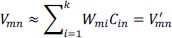
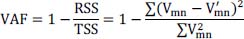
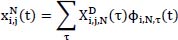
represents the signal in the i-th channel, j-th layer : denotes the wavelet packet coefficients, which indicate the projection weights of the signal onto the current wavelet basis; and ϕ(i,N,τ) (t) is the basis function for the N-th sub-band, used to capture the local time-frequency characteristics.

2-) Perform NMF decomposition:

where VjN represents the time-frequency signal matrix corresponding to the j-th layer and N-th sub-band. WjN is the synergy matrix that characterizes the features of different synergy patterns in the sub-band signal, and CjN is the temporal coefficient matrix, which indicates the time-varying contributions of the synergy patterns.

K-means clustering

The CMJ synergy modules from all 15 subjects were classified using a k-means clustering algorithm. Since VAF represents four cooperative modes, K was set to 4. To reduce the randomness introduced by the initial clustering centroids, the process was repeated 50 times using different initial centroids (Fan et al., 2024). The optimal number of clusters was determined using the Elbow Method, which identifies the inflection point in the error sum of squares relative to the number of clusters.

Statistical analyses

The Shapiro-Wilk test was used to assess the normality of data distribution. For normally distributed variables, paired sample t-tests were conducted to analyze pre- and post-intervention differences. For non-normally distributed variables, Mann-Whitney U tests were performed. Effect sizes were calculated using Cohen's d, with values of 0.2, 0.5, and 0.8 representing small, moderate, and large effects, respectively. All statistical analyses were conducted using MATLAB 2022a and GraphPad Prism 9.

RESULTS
Changes in CMJ performance

Both CMJ height (0.417 ± 0.066 vs 0.442 ± 0.068 m; P < 0.001, d = 0.45) and RSImod (0.51 ± 0.11 vs 0.56 ± 0.12; P < 0.001, d = 0.34) increased from pre- to post-BFR exercise (Figure 4).

Number of synergy modules across different time-frequency segments

The number of synergy modules observed before and after BFR training across the four frequency bands were as follows (Figure 5): i) 0-62.5 Hz: 4.00 ± 0.53 and 3.93 ± 0.45 (p = 0.726); ii) 62.5-125 Hz: 4.41 ± 0.49 and 4.52 ± 0.50 (p = 0.607); iii) 125-187.5 Hz: 4.55 ± 0.50 and 4.24 ± 0.57 (p = 0.068); and iv) 187.5-250 Hz: 4.38 ± 0.55 and 4.24 ± 0.50 (p = 0.460).

Muscle synergy analysis of CMJ

Muscle synergy analysis of synergy 1, which occurs during the Unweighting to Braking Phases of the CMJ, showed that the ankle dorsiflexor TA had the highest contribution both pre- and post-BFR training (weights = 0.78 and 0.81, respectively).

In Synergy 2, which is active during the Braking to Propulsion Phases of the CMJ, the ankle plantar flexor GL was the primary contributor (weights = 0.79 for both pre-and post-BFR), with a significant increase in GM activation post-training (weight Δ% = 33%; p = 0.032).

Synergy 3, active during the Propulsion to Flight Phases, revealed that the knee extensors RF (weights = 0.41, 0.43), VL (weights = 0.42, 0.36), and VM (weights = 0.5, 0.46) were the main contributors. Significant differences were observed between Pre-and Post-BFR intervention for GM (weight Δ% = 57%; p = 0.013) and VM (weight Δ% = -8%, p = 0.039).

Synergy 4, active during the Flight-to-Landing Phase of CMJ, showed greater contributions from the knee flexors ST (weight = 0.34, 0.43) and BF (weight = 0.54, 0.51), as well as the hip extensor GM. A significant post-BFR exercise increase was noted for VL (weight Δ% = 92%, p = 0.024) (Figure 6).

Muscle synergy analysis of CMJ in the time-frequency domain

A comprehensive analysis of muscle activation weights across frequency bands revealed significant differences pre- and post-BFR exercise differences (Figure 7). In the 0-62.5 Hz frequency band: i) TA in synergy 1 showed a significant increase (weight Δ% = 10%, p = 0.04); ii) GM in synergy 2 exhibited a marked increase (weight Δ% = 238%, p = 0.0007); and iii) in synergy 4, BF (weight Δ% = 32%, p = 0.032) and GM (weight Δ% = 11%, p = 0.034) also showed significant increases.

In the 62.5-125 Hz frequency band, significant changes were found in synergy 3, with a decrease in VL activation weight (weight Δ% = -39%, p = 0.003) and an increase in GM activation weight (weight Δ% = 87%, p = 0.009).

In the 125-185 Hz frequency band, significant differences were found in: i) synergy 2, with a significant increase in GM activation (weight Δ% = 53%, p = 0.031); and ii) synergy 4, where VM showed a significant increase (weight Δ% = 100%, p = 0.007).

In the 187.5-250 Hz frequency band, significant differences were found in: i) synergy 1, where GM activation significantly decreased (weight Δ% = -81%, p = 0.002); and ii) synergy 3, where VL exhibited a decrease in activation weight (weight Δ% = -22%, p = 0.002), and TA demonstrated a significant increase (weight Δ% = 75%, p = 0.001).

DISCUSSION

This study is the first to investigate neuromuscular control strategies underlying the effects of BFR training on CMJ performance using muscle synergy analysis. By employing WPT-NMF, we analyzed how BFR squats influence muscle activation and synergy across different sEMG frequency bands during CMJ. Our main findings indicate that acute BFR exercise significantly enhances CMJ height and RSImod. Although the total number of synergy modules remained unchanged, time-frequency analysis revealed adaptive shifts in activation weights. The results support our hypothesis that acute BFR resistance exercise improves CMJ performance by modifying time-frequency muscle synergy patterns.

We observed that acute BFR squats exercise significantly improves CMJ performance, as evidenced by improved jump height and RSImod. This immediate enhancement may stem from BFR-induced hypoxia, which stimulates type III and IV afferent nerves, promoting greater motor unit recruitment and particularly of type II fibers (Yasuda et al., 2015). Likewise, BFR exercise increased muscle activation synergy in the EMG signal and enhanced the secretion of endogenous hormones (Yasuda et al., 2010). Under ischemic conditions, improved synchronization of high-threshold motor unit firing may reduce antagonist co-activation, thus augmenting force production during explosive movements (Fatela et al., 2016; Loenneke et al., 2011). Additionally, BFR training has been shown to induce post activation performance enhancement and improve horizontal explosiveness and strength (Wharemate, 2021). Collectively, these performance benefits are likely driven by neuromuscular adaptations resulting from metabolic stress.

One key finding was that the number of muscle synergy modules across different frequency bands after WPT-NMF decomposition did not change following the BFR squat exercise. This indicates that CMJ performance is governed by stable neuromuscular control strategies. The consistency in synergy number may reflect a preserved role of specific frequency bands in muscle coordination. For instance, six muscle synergies were identified during running, both before and after fatigue, with no significant changes in the number of synergy modules, though muscle weights and activation timing shifted (Xu et al., 2025). Similarly, the number of muscle synergy modules remained unchanged across varying slopes and running speeds in uphill and downhill running, despite differences in muscle activation weights (Saito et al., 2018). These findings, including ours, suggest that the CNS maintains a stable core control framework while flexibly adjusting activation patterns (i.e., activation timing or weights) to meet task demands, such as those induced by BFR.

This study showed that while the number of muscle synergy modules during CMJ remains stable following acute BFR exercise, notable shifts in muscle activation weights occurred - particularly during the braking-landing phases. The dominant contributions of the dorsiflexor TA (Synergy 1) and plantar flexor GL (Synergy 2) reflect the importance of ankle joint stability during the braking phase (Chang et al., 2008). Their unchanged activation weights post-BFR suggest that distal stiffness is more influenced by task demands than by metabolic stress. The reduced contribution of VM during propulsion (Synergy 3) and the redistribution of VL activity during landing (Synergy 4) reflect task-specific adaptations aimed at optimizing knee joint torque. Comparable compensatory activation patterns have been observed under fatigue, such as during rowing, where redistribution helps maintain performance (Turpin et al., 2011). Enhanced GM activation across transitional phases (Synergy 2/3) further supports the concept of proximal muscle potentiation following BFR-induced metabolic stress (Fatela et al., 2019). These findings suggest that BFR modulates neural drive within pre-existing synergy structures, rather than creating new coordination patterns, a principle congruent with hierarchical motor control frameworks (Ting et al., 2015). Overall, BFR exercise enhances short-term explosive performance by fine-tuning neural input within stable synergy frameworks.

This study is the first to reveal frequency band-specific adaptations in neuromuscular control following acute BFR exercise during CMJ, using time-frequency domain muscle synergy analysis. Post-intervention, significant shifts in muscle activation weights across various frequency bands suggest that the CNS modulates performance through frequency-dependent strategies. Lower-frequency components in the sEMG signals are generally linked to the recruitment of slow-twitch, synchronized motor units, influenced by factors such as motor unit conduction velocity and volume-conductor effects (Farina et al., 2014). BFR-induced vascular occlusion leads to metabolite accumulation, stimulating sympathetic activity and increasing plasma levels of norepinephrine, epinephrine, and growth hormone (Takarada et al., 2000). High occlusion pressures may further activate mechanoreceptors, enhancing sympathetic responses (Williamson et al., 1994). In this study, increased activation weights in lower-frequency bands f particularly in dorsiflexors and hip extensors suggest that BFR-induced metabolic stress and neurohormonal responses may enhance ankle dorsiflexion stability and improve coordination between hamstrings and gluteus muscles. This low-frequency synchronization likely supports more efficient intermuscular coordination, thereby contributing to the stability and performance improvements observed in CMJ.

Changes in the sEMG signal spectrum are closely related to the recruitment patterns of fast motor units, which are progressively recruited during high-intensity contractions and exhibit higher firing frequencies than slow motor units, particularly under increased force demands. In this study, the activation differences in VL and GM within the high-frequency band (62.5-185 Hz) suggest that BFR selectively facilitates fast-twitch fibers recruitment, likely driven by metabolic byproducts accumulation (e.g., H+, inorganic phosphate) - an effect supported by previous research (Fatela et al., 2019). The mid-to-high frequency response of GM may compensate for metabolic limitations in distal muscles by increasing hip joint torque (Pandy and Zajac, 1991), thereby improving knee extension efficiency. Additionally, external limb compression may reduce local muscle activation and muscle cross-sectional area beneath the cuff, potentially inhibiting muscle activation in these regions (Ellefsen et al., 2015). As a compensatory mechanism, increased GM activation may offset this inhibition, improving overall movement performance and neuromuscular efficiency.

During explosive or rapid contractions, motor neurons are recruited within a short time and discharge at high frequencies (over 200 Hz) to support quick force generation (Del Vecchio et al., 2019). In this study, muscle synergy analysis in the high-frequency band (187.5-250 Hz) revealed synchronized activation of GM, VL, and TA likely due to BFR-induced local hypoxia, which triggers high-frequency discharge in fast-twitch muscle fibers. A similar phenomenon was noted previously, where BFR training significantly increased initial firing frequency, average discharge rate, and normalized EMG amplitude, indicating higher motor unit activation levels under the same load (Olmos et al., 2024). These findings suggest that BFR may enhance high-frequency muscle discharges through local hypoxia and metabolic stress, thereby modulating motor unit recruitment patterns to support rapid force production. Overall, our findings reinforce the notion that the CNS adapts through hierarchical, frequency-specific regulation, with BFR influencing muscle synergy by altering time-frequency muscle coordination patterns.

Although this study revealed that BFR exercise can enhance lower limb power output and RSImod by influencing muscle synergy, several limitations should be acknowledged. Firstly, due to equipment constraints, sEMG data were collected from only eight lower-limb muscles. The selection and number of muscles analyzed can influence muscle synergy outcomes (Steele et al., 2013). Secondly, the absence of 3D motion capture limited our ability to assess kinematic variables and calculate joint torque parameters, resulting in a narrower analytical scope. Although this study explored the physiological mechanisms of BFR exercise through changes in muscle synergy patterns, it did not directly assess neural adaptations. Future research should consider incorporating neurophysiological tools, such as electroencephalography or functional magnetic resonance imaging, to provide deeper insights into how BFR exercise influences the neuromuscular system.

CONCLUSION

Acute BFR exercise significantly enhances CMJ height and RSImod. Although the number of muscle synergy modules did not change, notable shifts in muscle activation weights occurred during key CMJ phases. These findings suggest that the CNS enhanced CMJ performance by modulating activation parameters within existing synergy modules across different frequency bands, rather than altering the synergy architecture itself. This study also demonstrates the time-frequency dynamics of synergy modulation following acute BFR squat exercise, offering new insights into the intrinsic neural patterns underlying enhanced athletic performance.

ACKNOWLEDGEMENTS

This study was conducted in accordance with the current laws and regulations of China. This work was supported by the "Pioneer" and "Leading Goose" R&D Program of Zhejiang (Grant No. 2025C02044) and the Fundamental Research Funds for the Central Universities (Grant No. 2024JNPD003). The authors have no conflict of interest to declare. This study received no specific grants, fellowships, or materials gifts from any funding agency in the public, commercial, or not-for-profit sectors. The datasets generated during and/or analyzed during the current study are not publicly available but are available from the corresponding author who organized the study.

AUTHOR BIOGRAPHY
     
 
Chenxi Hu
 
Employment:
 
Degree: PhD Candidate
 
Research interests: Physical Conditioning; Neuromuscular Control Mechanism
  E-mail: hcx212324@163.com


Ning Du
 
Employment:
 
Degree: PhD Candidate
 
Research interests: Sports physiology; Physical Therapy
  E-mail: duning304@163.com


Yanfeng Li
 
Employment:
 
Degree: Master
 
Research interests: Physical Conditioning; Sports Physiology
  E-mail: liyanfeng2022@cupes.edu.cn


Olivier Girard
 
Employment:
 
Degree: PhD
 
Research interests: Fatigue mechanisms during high-intensity intermittent exercises performed under challenging environmental conditions (i.e. heat stress or hypoxia), with a special focus on (repeated) sprinting mechanics and underpinning neuromuscular factors.
  E-mail: olivier.girard@uwa.edu.au


Tao Mei
 
Employment:
 
Degree: PhD
 
Research interests: Individual differences in exercise health promotion; Phenotypes and mechanisms for the principles and mechanisms of precision fitness coaching programs
  E-mail: meitao@bsu.edu.cn


REFERENCES
Beltrame G., Scano A., Marino G., Peccati A., Molinari Tosatti L., Portinaro N. (2023) Recent developments in muscle synergy analysis in young people with neurodevelopmental diseases: A systematic review. Frontiers in Bioengineering and Biotechnology 11, 1145937.
Chang S., Hu C. (2025) Comparative effects of narrow vs. wide cuff blood flow restriction on muscle synergy dynamics: A time-frequency decomposition approach. Sensors (Basel) 25.
Chang Y.H., Roiz R.A., Auyang A.G. (2008) Intralimb compensation strategy depends on the nature of joint perturbation in human hopping. Journal of Biomechanics 41, 1832-1839.
Comfort, P., Jones, P.A. and McMahon, J.J. (2018) Performance assessment in strength and conditioning. Routledge.
Cornejo-Daza P.J., Sánchez-Valdepeñas J., Rodiles-Guerrero L., Boullosa D., León-Prados J.A., Wernbom M., Pareja-Blanco F. (2025) Training effects of traditional versus cluster set configuration with and without blood flow restriction. Medicine & Science in Sports & Exercise 57, 668-679.
Del Vecchio A., Negro F., Holobar A., Casolo A., Folland J.P., Felici F., Farina D. (2019) You are as fast as your motor neurons: Speed of recruitment and maximal discharge of motor neurons determine the maximal rate of force development in humans. The Journal of Physiology 597, 2445-2456.
Ellefsen S., Hammarström D., Strand T.A., Zacharoff E., Whist J.E., Rauk I., Nygaard H., Vegge G., Hanestadhaugen M., Wernbom M., Cumming K.T., Rønning R., Raastad T., Rønnestad B.R. (2015) Blood flow-restricted strength training displays high functional and biological efficacy in women: A within-subject comparison with high-load strength training. American Journal of Physiology - Regulatory, Integrative and Comparative Physiology 309, 767-779.
Enoka R.M. (2008) Comments on Point:Counterpoint: Spectral properties of the surface EMG can characterize/do not provide information about motor unit recruitment strategies and muscle fiber type. Journal of Applied Physiology 105, 1676.
Fan P., Yang Z., Wang T., Li J., Kim Y., Kim S. (2024) Neuromuscular control strategies in basketball shooting: Distance-dependent analysis of muscle synergies. Journal of Sports Science and Medicine 23, 571-580.
Farina D., Merletti R., Enoka R.M. (2014) The extraction of neural strategies from the surface EMG: An update. Journal of Applied Physiology 117, 1215-1230.
Fatela P., Mendonca G.V., Veloso A.P., Avela J., Mil-Homens P. (2019) Blood flow restriction alters motor unit behavior during resistance exercise. International Journal of Sports Medicine 40, 555-562.
Fatela P., Reis J.F., Mendonca G.V., Avela J., Mil-Homens P. (2016) Acute effects of exercise under different levels of blood-flow restriction on muscle activation and fatigue. European Journal of Applied Physiology 116, 985-995.
Goudriaan M., Papageorgiou E., Shuman B.R., Steele K.M., Dominici N., Van Campenhout A., Ortibus E., Molenaers G., Desloovere K. (2022) Muscle synergy structure and gait patterns in children with spastic cerebral palsy. Developmental Medicine & Child Neurology 64, 462-468.
Guo W., Kim Y., Wang J., Dong T., Tang X., Kim S. (2025) 60-second static stretching of lower limb muscles disrupts muscular performance and control in active male adults. Journal of Sports Science and Medicine 24, 195-204.
Healy R., Kenny I.C., Harrison A.J. (2018) Reactive strength index: A poor indicator of reactive strength?. International Journal of Sports Physiology and Performance 13, 802-809.
Jagim A.R., Schuler J., Szymanski E., Khurelbaatar C., Carpenter M., Fields J.B., Jones M.T. (2024) Acute responses of low-load resistance exercise with blood flow restriction. Journal of Functional Morphology and Kinesiology 9.
Kraemer W.J., Ratamess N.A. (2004) Fundamentals of resistance training: Progression and exercise prescription. Medicine & Science in Sports & Exercise 36, 674-688.
Loenneke J.P., Fahs C.A., Wilson J.M., Bemben M.G. (2011) Blood flow restriction: The metabolite/volume threshold theory. Medical Hypotheses 77, 748-752.
Loenneke J.P., Kim D., Fahs C.A., Thiebaud R.S., Abe T., Larson R.D., Bemben D.A., Bemben M.G. (2015) Effects of exercise with and without different degrees of blood flow restriction on torque and muscle activation. Muscle & Nerve 51, 713-721.
Lorenz D.S., Bailey L., Wilk K.E., Mangine R.E., Head P., Grindstaff T.L., Morrison S. (2021) Blood flow restriction training. Journal of Athletic Training 56, 937-944.
Matsunaga N., Aoki K., Kaneoka K. (2021) Comparison of modular control during side cutting before and after fatigue. Applied Bionics and Biomechanics.
McKay A. K. A., Stellingwerff T., Smith E. S., Martin D. T., Mujika I., Goosey-Tolfrey V. L., Sheppard J., Burke L. M. (2022) Defining Training and Performance Caliber: A Participant Classification Framework. International Journal of Sports Physiology and Performance 17, 317-331.
Oláh V., Třebický V., Maleček J., Michalička V., Wąsik J., Vágner M. (2025) Is countermovement jump height and one repetition maximum back squat associated with the peak force of a front kick with and without carried load?. Journal of Strength and Conditioning Research.
Olmos A.A., Montgomery T.R., Sears K.N., Roth B.L., Richardson L.D., Dinyer-McNeely T.K., Hammer S.M., Bergstrom H.C., Hill E.C., Succi P.J., Lubiak S., Trevino M.A. (2024) Blood flow restriction increases motor unit firing rates and input excitation of the biceps brachii during a moderate-load muscle action. Journal of Sports Sciences 42, 1891-1903.
Pandy M.G., Zajac F.E. (1991) Optimal muscular coordination strategies for jumping. Journal of Biomechanics 24, 1-10.
Patterson S.D., Hughes L., Warmington S., Burr J., Scott B.R., Owens J., Abe T., Nielsen J.L., Libardi C.A., Laurentino G., Neto G.R., Brandner C., Martin-Hernandez J., Loenneke J. (2019) Blood flow restriction exercise: Considerations of methodology, application, and safety. Frontiers in Physiology 10, 533.
Ramirez-Campillo R., García-Hermoso A., Moran J., Chaabene H., Negra Y., Scanlan A.T. (2022) The effects of plyometric jump training on physical fitness attributes in basketball players: A meta-analysis. Journal of Sport and Health Science 11, 656-670.
Saito A., Tomita A., Ando R., Watanabe K., Akima H. (2018) Muscle synergies are consistent across level and uphill treadmill running. Scientific Reports 8, 5979.
Steele K.M., Tresch M.C., Perreault E.J. (2013) The number and choice of muscles impact the results of muscle synergy analyses. Frontiers in Computational Neuroscience 7, 105.
Sun D., Yang T. (2023) Semi-squat exercises with varying levels of arterial occlusion pressure during blood flow restriction training induce a post-activation performance enhancement and improve vertical height jump in female football players. Journal of Sports Science and Medicine 22, 212-225.
Takarada Y., Takazawa H., Ishii N. (2000) Applications of vascular occlusion diminish disuse atrophy of knee extensor muscles. Medicine & Science in Sports & Exercise 32, 2035-2039.
Ting L.H., Chiel H.J., Trumbower R.D., Allen J.L., McKay J.L., Hackney M.E., Kesar T.M. (2015) Neuromechanical principles underlying movement modularity and their implications for rehabilitation. Neuron 86, 38-54.
Turpin N., Guével A., Durand S., Hug F. (2011) Fatigue-related adaptations in muscle coordination during a cyclic exercise in humans. The Journal of Experimental Biology 214, 3305-314.
Turpin N.A., Uriac S., Dalleau G. (2021) How to improve the muscle synergy analysis methodology?. European Journal of Applied Physiology 121, 1009-1025.
Vigotsky A.D., Halperin I., Lehman G.J., Trajano G.S., Vieira T.M. (2017) Interpreting signal amplitudes in surface electromyography studies in sport and rehabilitation sciences. Frontiers in Physiology 8, 985.
Wang J., Fu H., QiangZhang Zhang M., Fan Y. (2022) Effect of leg half-squat training with blood flow restriction under different external loads on strength and vertical jumping performance in well-trained volleyball players. Dose Response 20, 15593258221123673.
Wharemate, J. (2021) Blood flow restriction as a method to elicit post activation potentiation in trained female athletes. The University of Waikato.
Williamson J.W., Crandall C.G., Potts J.T., Raven P.B. (1994) Blood pressure responses to dynamic exercise with lower-body positive pressure. Medicine & Science in Sports & Exercise 26, 701-708.
Wortman R.J., Brown S.M., Savage-Elliott I., Finley Z.J., Mulcahey M.K. (2021) Blood flow restriction training for athletes: A systematic review. American Journal of Sports Medicine 49, 1938-1944.
Xie P., Chang Q., Zhang Y., Dong X., Yu J., Chen X. (2022) Estimation of time-frequency muscle synergy in wrist movements. Entropy (Basel) 24.
Xu Y., Yang Y., He S., Yang C., Zhang S., Fu W., Li L. (2025) Running-induced fatigue influences lower extremity muscle synergy and related biomechanics. Gait & Posture 119, 163-170.
Yasuda T., Fujita S., Ogasawara R., Sato Y., Abe T. (2010) Effects of low-intensity bench press training with restricted arm muscle blood flow on chest muscle hypertrophy: A pilot study. Clinical Physiology and Functional Imaging 30, 338-343.
Yasuda T., Loenneke J.P., Thiebaud R.S., Abe T. (2015) Effects of detraining after blood flow-restricted low-intensity concentric or eccentric training on muscle size and strength. Journal of Physiological Sciences 65, 139-144.
Bao Nguyen, T., Yano, S. and Kondo, T. (2017) Muscle synergy analysis in dart throwing. Annual International Conference of the IEEE Engineering in Medicine and Biology Society 2534-2537.


Back
|
PDF
|
Share