|
|
|
| |
| ABSTRACT |
|
Table tennis athletes are required to execute appropriate footwork moving to the best position to hit the ball, while the chasse-step and one-step are typically employed in table tennis. This study aims to examine the difference in joint angles, joint moments, joint contact forces, and activation of lower limb muscles during the stance of chasse-step and one-step. Eighteen male table tennis athletes volunteered to perform topspin forehand with chasse-step and one-step. An eight-camera motion capture system and instrumented force plate were used to record makers’ trajectories and ground reaction force, which was then used to calculate the kinematics and kinetics with Inverse Kinematics and Inverse Dynamics in OpenSim. Surface electromyography signals were measured to validate the musculoskeletal OpenSim modeling. Hip flexion angle and moment increased in the backward swing phase during the stance of one-step. Knee extension of the chasse-step increased more during the forward swing phase. Hip contact force increased in the anterior-posterior direction of one-step and the chasse-step in the medial-lateral direction. Key findings suggest that the chasse-step may increase the quality of footwork performance and prepare the next step but shows higher injury risk in knee joints. While the one-step may have faster performance for scoring and high injury risk in hip joint. The information may provide implications for athletes and coaches to improve athletic performance and develop specific footwork training schemes to prevent potential injuries. |
| Key words:
Table Tennis, Footwork, OpenSim, EMG, Musculoskeletal Modelling
|
Key
Points
- The current study reports the kinematic, kinetic, and muscle activation of lower limbs between chasse-step and one-step in table tennis.
- Chasse-step may allow better preparation for the next phase, while one-step may allow quicker point scoring. Information may suggest that two types of footwork are associated with increased injury risk in the hip and knee joints.
- OpenSim musculoskeletal modelling is reliable in predicting table tennis footwork muscle activities.
|
Table tennis is a complex racket sport involving movement coordination, power management, and balance control to produce athletic performance (Nikolakakis et al., 2020). To improve athletic abilities, table tennis athletes are required to master set-up, footwork, stroke, and several key technical skills. Fuchs et al., (2018) emphasized the significance of footwork in table tennis and mentioned that footwork analysis involved tracking and examining the athletes’ body movements toward the ball during the rally. During each movement, table tennis athletes should be capable to employ the proper footwork to reach a suitable position before stroke (Malagoli Lanzoni et al., 2010). Basic table tennis footwork includes one-step, chasse-step, slide-step, cross-step, and pivot-step (Lam et al., 2019; Malagoli Lanzoni, 2020). Among the notational match analysis, Malagoli Lanzoni et al., (2014) suggested that Asian athletes showed quicker footwork performance compared to Europeans. Malagoli Lanzoni, (2020) compared male and female table tennis athletes' competitions and found that males preferred one-step and pivot-step over females. Knowledge of footwork biomechanics would be of great interest and practical significance for table tennis athletes, coaches, and biomechanical researchers. Table tennis movement is associated with lower limb coordination patterns from the widely reported kinematic characteristics. He et al., (2021) demonstrated that elite athletes showed a larger angular changing rate of ankle dorsiflexion and range of motion, as well as plantarflexion, than medium-level athletes. During the forehand topspin loop, higher-level athletes had greater hip extension and internal rotation, as well as reduced internal rotation of the ankle and knee joint in the forward phase (Wang et al., 2018). Higher-level athletes exhibited reduced forefoot plantarflexion and abduction during the cross-step end phase (Shao et al., 2020). Our previous study about sex differences in chasse-step exhibited that the flexion angles of the hip and knee joints in males were larger throughout the movement phase, and the internal rotation angle of the hip joint was significant during the forward swing phase (Yang et al., 2021; 2022). Understanding lower limb biomechanics related to footwork could provide valuable insights for improving movement techniques and developing effective training programs. Kinetics of the lower limb may affect athletic performance and injury risk. Lower limbs, as the origin of energy, could transfer the optimal activation of each segment in the lower limb upward through the kinetic chain to improve stroke quality (Elliott, 2006). Taking tennis sport as an example of the kinetic chain, Ben Kibler (1995) studied tennis biomechanics and calculated that more than 50 percent of the total force was generated through the lower limbs, including lower leg, hip, and torso. In the whole kinetic chain of the tennis serve, the lower limb and core provide a stable foundation, and the coordinated movements generate the most effective force production to hit the ball (Saini et al., 2020). While exploring tennis-related injuries, significant stress on joints and the spine were similarly observed in table tennis. In addition, due to the large number of muscle impulses required for execution in the case of techniques in tennis, the situation was similar to table tennis (Mocanu et al., 2020). Quantifying kinetic and muscle activation characteristics could provide biomechanical insights to help table tennis athletes and coaches optimize footwork strategies. Previous table tennis-related studies mainly focused on kinematic analysis, but little information was reported about joint contact forces and muscle activations of table tennis footwork, which may affect performance and injuries (Iino, 2018; He et al., 2022; Yang et al., 2022). Thus, this study aims to investigate the joint kinematics, joint kinetics, joint contact forces, and muscle activations between table tennis chasse-step and one-step footwork. It is hypothesized that two different footwork would show significant differences in several biomechanical characteristics of the lower limb. The information would assist table tennis coaches and athletes in understanding the biomechanical mechanisms of footwork performance and developing training schemes to prevent potential injuries. ParticipantsEighteen elite Chinese male table tennis athletes (National Level I) volunteered to join this study (Table 1). All participants had no history of lower limb pain or disorders within the past two years and were free from injury for at least six months before this study. All participants held the racket with the right hand employed for a match or competition, defined as dominant side. All participants were informed of the benefits and risks of the study before signing consent. This research was approved by the Human Ethics Committee from the research institute at Ningbo University (RAGH20211009).
Experimental ProceduresAn eight-camera motion capture Vicon system (Oxford Metrics Ltd., Oxford, UK) was used to capture the marker trajectories with a frequency of 200Hz. An in-ground instrumented force plate (AMTI, Watertown, MA, US) was utilized to record the ground reaction force with a frequency of 1000 Hz. A 52-marker set (diameter: 14 mm) was used for all participants during the experiment. The markers were attached to the left and right upper and lower limb, which locations included: the shoulder, distal joint of humerus, radius and ulna, the proximal joint of the second and fifth phalanx, iliac spine, condyle, malleolus, first and fifth metatarsal heads, distal joint of the first and second toe, as well as tracking clusters attached to the elbow, wrist, thigh, shank, and heel (Figure 1i). Surface electromyography (EMG) signals were recorded via an EMG system (Delsys, Boston, MA, United States) for muscle activities. Surface electrodes were attached to the lower limb (Figure 1_ii), including the rectus femoris (RF), bicep femoris lateral (BF), vastus lateralis (VL), vastus medialis (VM), tibia anterior (TA), gastrocnemius medial (GM), gastrocnemius lateral (GL). All participants were required to jog for 15 mins on the treadmill. After a warm-up and experimental environment familiarization, the electrodes were attached to the corresponding muscles' skin surface (belly), which was wiped with alcohol to reduce impedance (Wang et al., 2018). To obtain the maximal value of the muscle activity, the athletes performed two maximum voluntary contractions (MVC) tests with isometric contractions. Experimental tests were conducted in a motion capture lab facilitated with eight Vicon cameras and an in-ground AMTI force plate. All participants held the same table tennis racket and wore the same table tennis shoes. After a 15-minute classical multi-ball training with chasse-step and one-step, all athletes were asked to hit the ball with two different footwork, including two shots in chasse-step and one shot in one-step. For all topspin forehand serves by the coach, each participant, using a chasse-step from the first (Figure 2ia) and final impact zones (Figure 2ib), and one-step from the final impact zone separately, hit the ball onto the diagonal court, which was the target zone (Figure 2_i_c). The table tennis ball employed for this study was the same as that used in most competitions, with the brand information (D40+, Double Happiness Sports Company, Shanghai, China). The smoothness of motion and quality of strokes were judged by the professional coach who served the ball during the experiment. Data of marker trajectories and ground reaction force from one static and four successful trials, including lower limb EMG signals, were synchronously collected during the foot landing on the force plate, which is the last shot in the final impact zone.
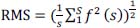
Data processing and musculoskeletal modelingThis study mainly analyzed the stance landing on the force plate of chasse-step and one-step. As per the arm movement, the stance was divided into the backward swing phase (BP) and forward swing phase (FP), as previously validated in our recent study (Yang et al., 2021). After standardization, participants entered the BP (Figure 2_ii_b) when landing on the force plate with footwork, about 30% of the whole stance was the end of BP (Figure 2_iv_c), followed by the FP (Figure 2_ii_d), and 60% of the stance was the end of FP (Figure 2_ii_e), and then returned to the ready position (Figure 2_ii_a). This study employed a previously validated OpenSim Musculoskeletal model (v4.2) for data processing as per the established pipelines (Hamner and Delp, 2013; Seth et al., 2011). The model was firstly scaled following the static markers position of each participant to achieve an anthropometric-match athlete-specific model. To reduce the error, the Inverse Kinematics (IK) with weighted markers was taken to calculate the joint angles. The joint angles were then used in an Inverse Dynamic (ID) algorithm to compute the joint moments. The Static Optimization algorithm analyzed the muscle activation and muscle forces. The muscle activation was compared against the EMG muscle activities to validate the musculoskeletal modeling in this study. The computed muscle forces during the footwork stance started to end with the platform's force for muscular contribution analysis. The surface EMG signals were filtered by bandpass (10-500Hz, FTT filter) with root mean square (RMS). By the two maximum voluntary contractions (MVC) tests, the highest value of the EMG envelope was retained as the reference. The same method was used to determine the EMG peak amplitude for chasse-step and one-step. The EMG amplitude was averaged for each muscle, followed by the footwork test. As in Equation 1, the root mean square (RMS) calculation algorithm was used for MVC and table tennis footwork trials in the Delsys EMG work Analysis software (S represents window length (points), and f(s) represents data in the window) (Klyne et al., 2012).
 |
|
(1) |
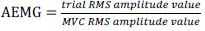
Model simulated activation was reported on a scale from zero to one from Opensim, with zero representing non-activation and one representing maximum activation (Hamner et al., 2010; Hamner and Delp, 2013; Rajagopal et al., 2016). As Equation 2, EMG signals activities of two different footwork were normalized to a zero-one scale (Schmid et al., 2010).
 |
|
(2) |
Statistical analysisThe statistical analysis included the joint kinematics, joint moments, and joint contact forces of the right lower limb. The normality of data (Shapiro-Wilk test) was checked before statistical analysis. Independent sample t-tests were taken for each dependent variable between the chasse-step and one-step. The open-source Statistical Parametric Mapping 1D package (SPM 1D) was utilized for statistical analysis. All extracted data were analyzed in MATLAB R2019a (The MathWorks, MA, United States), with a significance threshold 0.05.
Hip jointAs outlined in Figure 3i, hip joint angles between the chasse-step and one-step showed significant differences in the three-dimensional planes. At the hip joint during one-step, flexion increased at 0% - 5% (p = 0.045) and 59%-60% (p = 0.049) (Figure 3ia), while abduction increased from 15% to 50% (p < 0.001) (Figure 3ib). In addition, internal rotation increased at 20%-25% (p = 0.0045) (Figure 3ic). Figure 3ii shows the comparison of hip joint moments between the chasse-step and the one-step. Chasse-step increased more extension moment at 6% - 10% (p = 0.020) and more flexion moment at 62% - 68% (p = 0.0014), 85% - 95% (p < 0.001), 98% - 100% (p = 0.042) than one-step (Figure 3iia). Adduction moment increased at 50% - 60% (p < 0.001) and 78% - 83% (p = 0.001) during the stance of one-step (Figure 3iib). Increased external rotation moment of one-step was observed across stance at 55-62% (p = 0.007) and 78%-81% (p = 0.033) (Figure 3iic). Hip joint contact forces were reported in Figure 3iii. The contact force of the hip joint increased in the ant-post direction at 15% - 35% (p < 0.001) and 53% - 85% (p < 0.001) during one-step (Figure 3iiia). In the med-lat direction, the contact force of the chasse-step increased across stance at 0-2% (p = 0.049), 8% - 12% (p = 0.004), and 20% - 28% (p < 0.001), while one-step increased at 57% - 63% (p < 0.001) (Figure 3iiib). Furthermore, the contact force of the hip joint increased in the sup-inf direction at 35% - 48% (p < 0.001) during the stance of the chasse-step and 55% - 75% (p < 0.001) during the stance of the one-step (Figure 3iiic).
Knee jointFigure 4 shows the comparison of knee joint angles between the chasse-step and the one-step. The knee joint angle of one-step increased flexion from 40% to 85% (p < 0.001) and from 99% to 100% (p = 0.049) (Figure 4a). However, there were no significant differences observed in knee joint angles in the frontal and transverse planes, respectively (Figure 4b, Figure 4c).
As shown in Figure S1 of supplementary, the extension moment increased at 12% - 25% (p < 0.001) of the chasse-step, 40% - 75% (p < 0.001) of the one-step (Figure S1_a). Adduction moment increased at 55% - 63% (p = 0.002) during one-step (Figure S1b). During chasse-step, the knee external rotation moment increased more at 28% - 35% (p < 0.001) than one-step (Figure S1c). Knee contact forces were shown in Figure S2 during the chasse-step and one-step. The contact force increased in the ant-post-direction at 0-1% (p = 0.049) during stance of chasse- step, but 45% - 75% (p < 0.001) during stance of one-step (Figure S2a). The knee contact force increased during chasse-step stance at 8%-10% (p = 0.042) in the med-lat direction (Figure S2b). In the sup-inf direction, the contact force of chasse-step was increased across stance at 6% - 9% (p = 0.032) and 18% - 22% (p = 0.05). However, the contact force in the sup-inf direction of one-step increased during 55% - 75% (p < 0.001) (Figure S2c).
Ankle jointThe ankle angles and moments were showed in Figure S3. One-step had more dorsiflexion than chasse-step at 45%-68% (p = 0.002) (Figure S3_a) and showed more plantarflexion moment at 55% - 72% (p < 0.001) (Figure S3_b). As shown in Figure S4, ankle contact force of chasse-step increased at 2% - 6% (p = 0.025) in the ant-post direction and 2% - 4% (p = 0.026) in the med-inf direction. Additionally, ankle contact force increased at 57% - 72% (p < 0.001) in the ant-post direction and 50% - 70% (p < 0.001) in the med-inf direction during stance of one-step (Figure S4_a, Figure S4_b). The contact force of ankle joint increased at 2% - 5% (p = 0.021) with chasse-step, and 58% - 73% (p < 0.001) with one-step in the sup-inf direction (Figure S4_c).
EMGThe EMG signals from the experiments were converted to activation, from 0 (no activation) to 1 (full activation). Figure 5 presents seven lower limb muscles of the EMG amplitude calculated for the chasse-step (Figure 5a) and one-step (Figure 5b). Most muscle activities were similar between OpenSim modelling and EMG signals. RF was moderated to strongly activated during the stance of one-step. Both VM and VL EMG activity levels measured were smaller and statistically different between the two table tennis steps. BF and GL showed 60% activation with chasse-step and one-step. GM exhibited a greater level of activity during the chasse-step. The one-step EMG amplitude of TA was measured to be greater than the chasse-step.
This study aimed to compare the kinematics and kinetics of the lower limb and discuss how muscle forces affect the dynamics of the lower extremity between chasse-step and one-step footwork in table tennis. The findings of this study supported the hypothesis that joint angles, moments, and contact forces differed significantly between different footwork in table tennis. Specifically, hip joint flexion angle and moment increased in the backward swing phase of one-step. The knee joint extension angle increased during the forward swing phase in the chasse-step, but knee flexion angle increased in the one-step during the same swing phase. In the ant-post direction, hip joint contact force in one-step increased during the whole phase. In addition, the contact force of the hip joint increased during the chasse-step in the med-lat direction. There are no significant results observed in the ankle joint, thus not discussed in the current section but reported in the Appendix. After the end of the forward swing movement based on the z-component of displacement (Bańkosz and Winiarski, 2017), it was found that athletes had movements from back to the ready position with angles and moments defining this phase as a follow-swing phase. One-stepOne-step in table tennis was usually employed when the opponent quickly played the ball and spent a shorter time going to the right position to stroke the ball (Malagoli Lanzoni et al., 2010). Increased hip flexion angle and moment in the backward swing phase and increased knee flexion angle in the forward swing phase were observed in one-step, which may suggest that one-step may be used with insufficient time for stroke, so table tennis athletes employed the hip and surrounding muscles to stabilize the body for stroke return. A stable and strong lower limb foundation is crucial for precise whole-body movements in table tennis, which was highlighted that the movement of the hip joint played a crucial role in generating and transferring energy during table tennis (Qian et al., 2016). The hip provided dynamic stability for the whole kinetic chain in the process of table tennis movement (Gu et al., 2019; Wang et al., 2018). In addition, knee joint flexion in racket sports athletes is essential for maintaining precise stability (Lees, 2003). However, from the table tennis review of injuries, the high involvement of the hip and knee joints in table tennis made it a primary site of lower limb injury (Ferrandez et al., 2021; He et al., 2022). This key finding could be concluded that the one-step employed the hip and knee to keep stability, which may also hint at a higher injury risk. As an open chain of motion, table tennis was designed to accelerate the distal segment (Iino and Kojima, 2011). The greater contact force in the ant-post direction of the hip joint may allow athletes to use the dominant hand for forwarding to hit the ball returning the opponent. Similarly, an increase in the forward component of certain joint ranges of motion facilitated the impact-hitting force, which reported that the increased forward bending might improve the scoring rates (Bańkosz and Winiarski, 2018b). The generation of axial rotational torque in the pelvis on the dominant side was crucial for achieving high horizontal racket speed (Iino, 2018). The pelvis's backward tilting torque contributed to the acceleration of the racket speed generation, which may suggest that the one-step was typically employed to improve the faster performance for directly winning the point.
Chasse-stepThe hip contact force in the med-lat direction increased during the chasse-step, which may infer that the chasse-step used side direction movement moving to the appropriate position for preparation for the next stroke. Malagoli Lanzoni et al. (2014) reported that the chasse-step was used to execute a wide range of defensive and offensive strokes through side movements via comparing different footwork. Zhou et al. (2021) noted that the medial and lateral ground reaction force of the chasse-step was significantly larger than the cross-step. In the comparison between the long chasse-step and the short chasse-step, the short chasse-step was more conducive to efficiently transitioning to the next phase (Yu et al., 2019). Thus, it might be summarized that the chasse-step among table tennis footwork is related to maintaining the next strokes. Chasse-step could cover a medium distance and play harder stroke return (Malagoli Lanzoni et al., 2010). The knee extension angle was significantly increased during the forward swing phase in the chasse-step, which may be related to improving movement quality. Table tennis requires athletes to maintain a semi-bent knee position at an angle of 90 degrees or more over an extended period (Rajabi et al., 2012). High-level table tennis athletes could utilize the knee joint more efficiently to facilitate upper limbs than low-level athletes (Bańkosz and Winiarski, 2018a). By comparing the chasse-step between males and females, it was found that males with better stroke skills exhibited a greater range of motion in the knee joint during the forward swing phase but increased injury risk (Yang et al., 2021). Knowledge of the knee extension range in the dominant limb to perform better stroke might suggest that the chasse-step in table tennis could be stretched out to produce a stroke of high quality, but high loading profiles would be paid attention to reduce the risk of injury in the knee joint.
EMG validationRecent table tennis muscle activity research was more about upper limbs (Meghdadi et al., 2019), strokes (Yoichi et al., 2022), and athletic levels (Wang et al., 2018). Few studies focused on analysing muscle activation and forces via musculoskeletal modelling of table tennis footwork (He et al., 2022). The muscle activations obtained during the chasse-step and one-step from the static optimization in OpenSim simulation were similar to the EMG activity from experiments, indicating the reliability and validity of findings in this study (Hamner and Delp, 2013; Rajagopal et al., 2016). Considering the consistency of OpenSim musculoskeletal modeling (Delp et al., 1990, 2007; Seth et al., 2018) with the computed muscle control (CMC) and static optimization (SO) algorithm, the muscle excitations and forces were estimated (Thelen et al., 2003) by considering muscle activation (Zajac, 1989). The vastus lateralis, biceps femoris, gastrocnemius medialis, tibialis anterior, and other lower limb muscles were simulated to match the EMG activities, similar to running in a previous research (Hamner and Delp, 2013). Findings may indicate that the OpenSim model in this study could be utilized to understand the muscular contribution in table tennis footwork.
LimitationsAs a limitation of this study, the realistic scenarios might be difficult to duplicate considering the lab-based experimental environment and there is no robot machine used to serve the ball, which may explain the differences between the OpenSim modeling and EMG signals. More information about different muscle activation patterns, such as coordination (Kainz et al., 2024; Uhlrich et al., 2022), is needed to analyze and discuss different footwork. Furthermore, the upper extremity was not investigated in the musculoskeletal biomechanical analysis of table tennis footwork, and future research shall consider the upper and lower extremities combined to understand the footwork drive for power generation.
The study investigated the lower extremity biomechanics of chasse-step and one-step footwork in elite table tennis athletes. The key findings may suggest that one-step is associated with faster performance for a direct score while chasse-step is associated with performing high-quality strokes and efficient preparation for the next phase. However, the chasse-step is associated with risk of knee injury while the one-step is related to the hip and knee joint. Further, the OpenSim musculoskeletal modeling is reliable in table tennis footwork to verify the muscle activation against the EMG activities to understand muscular contribution. The knowledge of biomechanical loading profiles and muscular contribution may assist coaches in developing evidence-based training schemes and provide theoretical references for the scientific training of elite table tennis athletes for injury prevention and footwork performance.
| ACKNOWLEDGEMENTS |
The authors thank the valuable contribution of all the athletes and researchers who participated in this study. This study was supported by the National Natural Science Foundation of China (12202216), Ningbo Natural Science Foundation (2023J128), and the Mechanics Interdisciplinary Fund for Outstanding Young Scholars of Ningbo University (GC2024006), and K.W. Wong Magna Fund in Ningbo University. The experiments comply with the current laws of the country in which they were performed. The authors have no conflict of interest to declare. The datasets generated during and/or analyzed during the current study are not publicly available but are available from the corresponding author who was an organizer of the study. |
|
| AUTHOR BIOGRAPHY |
|
 |
Xiaoyi Yang |
| Employment: Auckland Bioengineering Institute, The University of Auckland, New Zealand |
| Degree: PhD Candidate |
| Research interests: Musculoskeletal Biomechanics |
| E-mail: xyan725@aucklanduni.ac.nz |
| |
 |
Yuming Wang |
| Employment: Faculty of Sports Science, Ningbo University |
| Degree: Master Student |
| Research interests: Sport Biomechanics |
| E-mail: wangyuming69@aliyun.com |
| |
 |
Qichang Mei |
| Employment: Associate Professor, Ningbo University |
| Degree: PhD |
| Research interests: Sport injury, Musculoskeletal modelling, Footwear Biomechanics |
| E-mail: meiqichang@outlook.com |
| |
 |
Shirui Shao |
| Employment: Faculty of Sports Science, Ningbo University, China |
| Degree: PhD Student |
| Research interests: Sport Biomechanics |
| E-mail: shaoshirui@nbu.edu.cn |
| |
 |
Yaodong Gu |
| Employment: Professor, Dean, Ningbo University |
| Degree: PhD |
| Research interests: Lower extremity biomechanics, Foot and footwear biomechanics |
| E-mail: guyaodong@hotmail.com |
| |
 |
Justin Fernandez |
| Employment: Professor, Auckland Bioengineering Institute, The University of Auckland, New Zealand |
| Degree: PhD |
| Research interests: Musculoskeletal Biomechanics |
| E-mail: j.fernandez@auckland.ac.nz |
| |
|
| |
| REFERENCES |
 Bańkosz Z., Winiarski S. (2017) The kinematics of table tennis racquet: differences between topspin strokes. The Journal of Sports Medicine and Physical Fitness 57. Crossref |
Bańkosz Z., Winiarski S. (2018a) Correlations between angular velocities in selected joints and velocity of table tennis racket during topspin Forehand and Backhand. Journal of Sports Science and Medicine 17, 330-338. Pubmed |
Bańkosz Z., Winiarski S. (2018b) The Evaluation of Changes of Angles in Selected Joints During Topspin Forehand in Table Tennis. Motor Control 22, 314-337. Crossref |
Ben Kibler W. (1995) Biomechanical Analysis of the Shoulder During Tennis Activities. Clinics in Sports Medicine 14, 79-85. Crossref |
Delp S L., Loan J.P., Hoy M.G., Zajac F.E., Topp E.L., Rosen J.M. (1990) An Interactive Graphics-Based Model of the Lower Extremity to Study Orthopaedic Surgical Procedures. IEEE Transactions on Biomedical Engineering 37, 757-767. Crossref |
Delp S.L., Anderson F.C., Arnold A.S., Loan P., Habib A., John C.T., Guendelman E., Thelen D.G. (2007) OpenSim: Open-source software to create and analyze dynamic simulations of movement. IEEE Transactions on Biomedical Engineering 54, 1940-1950. Crossref |
Elliott B. (2006) Biomechanics and tennis. British Journal of Sports Medicine 40, 392-396. Crossref |
Ferrandez C., Marsan T., Poulet Y., Rouch P., Thoreux P., Sauret C. (2021) Physiology, biomechanics and injuries in table tennis: A systematic review. Science and Sports 36, 95-104. Crossref |
Fuchs M., Liu R., Malagoli Lanzoni I., Munivrana G., Straub G., Tamaki S., Yoshida K., Zhang H., Lames M. (2018) Table tennis match analysis: a review. Journal of Sports Sciences 36, 2653-2662. Crossref |
Gordon D., Robertson E., Winter D.A. (1980) Mechanical energy generation, absorption and transfer amongst segments during walking. Journal of Biomechanics 13, 845-854. Crossref |
Gu Y., Yu C., Shao S., Baker J. S. (2019) Effects of table tennis multi-ball training on dynamic posture control. PeerJ 6, e6262. Crossref |
Hamner S.R., Delp S.L. (2013) Muscle contributions to fore-aft and vertical body mass center accelerations over a range of running speeds. Journal of Biomechanics 46, 780-787. Crossref |
Hamner S.R., Seth A., Delp S.L. (2010) Muscle contributions to propulsion and support during running. Journal of Biomechanics 43, 2709-2716. Crossref |
He Y., Fekete G., Sun D., Baker J.S., Shao S., Gu Y. (2022) Lower Limb Biomechanics during the Topspin Forehand in Table Tennis: A Systemic Review. Bioengineering 9, 1-16. Crossref |
He Y., Lyu X., Sun D., Baker J.S., Gu Y. (2021) The kinematic analysis of the lower limb during topspin forehand loop between different level table tennis athletes. PeerJ 9. Crossref |
Iino Y. (2018) Hip joint kinetics in the table tennis topspin forehand : relationship to racket velocity. Journal of Sports Sciences 36, 834-842. Crossref |
Iino Y., Kojima T. (2011) Kinetics of the upper limb during table tennis topspin forehands in advanced and intermediate players. 10, 361-377. Crossref |
Kainz H., Koller W., Wallnöfer E., Bader T.R., Mindler G.T., Kranzl A. (2024) A framework based on subject-specific musculoskeletal models and Monte Carlo simulations to personalize muscle coordination retraining. Scientific Reports 14, 1-13. Crossref |
Klyne D.M., Keays S.L., Bullock-Saxton J. E., Newcombe P.A. (2012) The effect of anterior cruciate ligament rupture on the timing and amplitude of gastrocnemius muscle activation: A study of alterations in EMG measures and their relationship to knee joint stability. Journal of Electromyography and Kinesiology 22, 446-455. Crossref |
Lam W.K., Fan J.X., Zheng Y., Lee W. C.C. (2019) Joint and plantar loading in table tennis topspin forehand with different footwork. European Journal of Sport Science 19, 471-479. Crossref |
Lees A. (2003) Science and the major racket sports: A review. Journal of Sports Sciences 21, 707-732. Crossref |
Malagoli Lanzoni I. (2020) Footwork technique used in elite table tennis matches. International Journal of Racket Sports Science 1, 44-48. Crossref |
Malagoli Lanzoni I., Di Michele R., Merni F. (2014) A notational analysis of shot characteristics in top-level table tennis players. European Journal of Sport Science 14, 309-317. Crossref |
Malagoli Lanzoni I., Lobietti R., Merni F. (2010) Footwork in Relationship with Strokes and Efficacy during the 29 th Olympic Games Table Tennis Final. International Journal of Table Tennis Sciences 6, 60-63. |
Meghdadi N., Yalfani A., Minoonejad H. (2019) Electromyographic analysis of shoulder girdle muscle activation while performing a forehand topspin in elite table tennis athletes with and without shoulder impingement syndrome. Journal of Shoulder and Elbow Surgery 28, 1537-1545. Crossref |
Mocanu M.D., Mereuţă C., Iordan D.A. (2020) Injuries resulting from practicing performance sports in table tennis and tennis. The Annals of “Dunarea de Jos” University of Galati Fascicle XV Physical Education and Sport Management 2, 12-23. Crossref |
Nikolakakis A., Mavridis G., Gourgoulis V., Pilianidis T., Rokka S. (2020) Effect of an intervention program that uses elastic bands on the improvement of the forehand topspin stroke in young table tennis Athletes. Journal of Physical Education and Sport 20, 2189-2195. Crossref |
Qian J., Zhang Y., Baker J. S., Gu Y. (2016) Effects of performance level on lower limb kinematics during table tennis forehand loop. Acta of Bioengineering and Biomechanics 18, 149-155. Crossref |
Rajabi R., Johnson G.M., Alizadeh M.H., Meghdadi N. (2012) Radiographic knee osteoarthritis in ex-elite table tennis players. BMC Musculoskeletal Disorders 13, 2-7. Crossref |
Rajagopal A., Dembia C.L., DeMers M.S., Delp D.D., Hicks J.L., Delp S.L. (2016) Full-Body Musculoskeletal Model for Muscle-Driven Simulation of Human Gait. IEEE Transactions on Biomedical Engineering 63, 2068-2079. Crossref |
Saini S.S., Shah S.S., Curtis A.S. (2020) Scapular Dyskinesis and the Kinetic Chain: Recognizing Dysfunction and Treating Injury in the Tennis Athlete. Current Reviews in Musculoskeletal Medicine 13, 748-756. Crossref |
Schmid S., Moffat M., Gutierrez G.M. (2010) Effect of knee joint cooling on the electromyographic activity of lower extremity muscles during a plyometric exercise. Journal of Electromyography and Kinesiology 20, 1075-1081. Crossref |
Seth A., Hicks J.L., Uchida T.K., Habib A., Dembia C.L., Dunne J.J., Ong C.F., DeMers M.S., Rajagopal A., Millard M., Hamner S.R., Arnold E.M., Yong J.R., Lakshmikanth S.K., Sherman M.A., Ku J.P., Delp S.L. (2018) OpenSim: Simulating musculoskeletal dynamics and neuromuscular control to study human and animal movement. Plos Computational Biology 14, e1006223. Crossref |
Seth A., Sherman M., Reinbolt J.A., Delp S. L. (2011) OpenSim: A musculoskeletal modeling and simulation framework for in silico investigations and exchange. Procedia IUTAM 2, 212-232. Crossref |
Shao S., Yu C., Song Y., Baker J.S., Ugbolue U.C., Lanzoni I.M., Gu Y. (2020) Mechanical character of lower limb for table tennis cross step maneuver. International Journal of Sports Science and Coaching 15, 552-561. Crossref |
Thelen D.G., Anderson F.C., Delp S.L. (2003) Generating dynamic simulations of movement using computed muscle control. Journal of Biomechanics 36, 321-328. Crossref |
Uhlrich S.D., Jackson R.W., Seth A., Kolesar J.A., Delp S.L. (2022) Muscle coordination retraining inspired by musculoskeletal simulations reduces knee contact force. Scientific Reports 12, 1-13. Crossref |
Wang M., Fu L., Gu Y., Mei Q., Fu F., Fernandez J. (2018) Comparative Study of Kinematics and Muscle Activity between Elite and Amateur Table Tennis Players during Topspin Loop Against Backspin Movements. Journal of Human Kinetics 64, 25-33. Crossref |
Yang X., He Y., Shao S., Baker J. S., István B., Gu Y. (2021) Gender differences in kinematic analysis of the lower limbs during the chasse step in table tennis athletes. Healthcare (Switzerland) 9, 1-13. Crossref |
Yang X., Mei Q., Shao S., Gu W., He Y., Zhu R., Gu Y. (2022) Understanding Sex-Based Kinematic and Kinetic Differences of Chasse-Step in Elite Table Tennis Athletes. Bioengineering 9, 1-10. Crossref |
Yoichi I., Shinsuke Y., Senshi F. (2022) Validation of lower limb muscle activation estimated using musculoskeletal modeling against electromyography in the table tennis topspin forehand and backhand. International Journal of Racket Sports Science 4, 1-10. Crossref |
Yu C., Shao S., Awrejcewicz J., Baker J.S., Gu Y. (2019) Lower Limb Maneuver Investigation of Chasse Steps Among Male Elite Table Tennis Players. Medicina 55, 97. Crossref |
Zajac F.E. (1989) Muscle and tendon: properties, models, scaling, and application to biomechanics and motor control. Critical Reviews in Biomedical Engineering 17, 359-411. Pubmed |
Zhou H., He Y., Yang X., Ren F., Ugbolue U.C., Gu Y. (2021) Comparison of kinetic characteristics of footwork during stroke in table Tennis: Cross-step and chasse step. Journal of Visualized Experiments 172. Crossref |
|
| |
|
|
|
|